Final project
Teams: 3-4 people
Report Due Date: 11:59pm Monday, April 25th (the night before the first set of presentations)
Deliverables:
Folder containing all the code files (no need to include data) used for project. Include a README.txt that briefly describes the functions/purposes of each file and the order you run them in.
Prepare a report that addresses all the major questions (3 for undergraduate section and 4 for graduate section). The report should be focused on detailing the methods and the exploration of the dataset. Please include labeled figures to better visualize the results and data.
Prepare a 10-15 minute presentation that summarizes the method used and the results.
Introduction:
Your collaborators are working on protocols to produce engineered mouse cardiomyocytes (CM) in vitro from fibroblasts. They have generated single-cell RNA-seq data and they are hoping you can help provide detailed hypotheses about the status of their cells and about potential protocol improvements. Their single-cell data are provided in the file folder direct_conversion_data (raw counts). These data are in fact from Stone et al. Batched 1, 2, 3, 4, 5, and 6 represent timepoints -1, 1, 2, 3, 7, and 14. We have subsampled the data to 5,000 cells total, which we hope will simplify analysis for people with less RAM. If you want and you have sufficient computational power, you can also use the entire Stone dataset in direct_conversion_data_full.
Major Questions:
How pure are your engineered cardiomyocytes?
For estimating the success of engineered cardiomyocyte from Stone et al (similarity of expression profiles to cardiac muscle cells or fibroblast), perform in silico validation of the expression profiles with reference dataset sampled from Tabula Muris. Develop a method to perform comparison between the engineered CMs and reference dataset. - You may choose to use pySCN to perform the assessment.
How mature are your engineered cardiomyocytes?
Heart is one of the earliest organs to develop during embryogenesis. In mice, as early as embryonic day E7.0-7.5, convergence of lateral mesoderm domain called first heart field begins to beat. Cardiomyocytes rapidly proliferate up until E14, and then start maturation after.
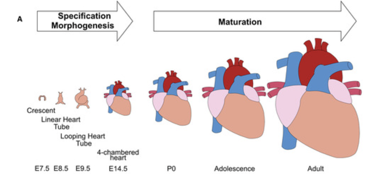
Conceptual scheme of maturation phase of heart development. Guo et al, 2020
Some of the major hallmarks of cardiomyocyte maturation include cell cycle arrest, multinucleation, growth by hypertrophy, transition to oxidative metabolism, and expression of mature sarcomeric contractile protein isoforms. Padula et al, 2021 provides a review of the transcriptional regulations of cardiomyocyte maturation and the TF genes associated with CM maturation phenotypes.
To computationally estimate the maturity of engineered CM’s across different data sources, Kannan et al constructed a reference scRNA-seq resource for CM maturation by isolating CM cells from Myh6-Cre; mTmG (aMHC x mTmG) mice across 12 time points of CM maturation.
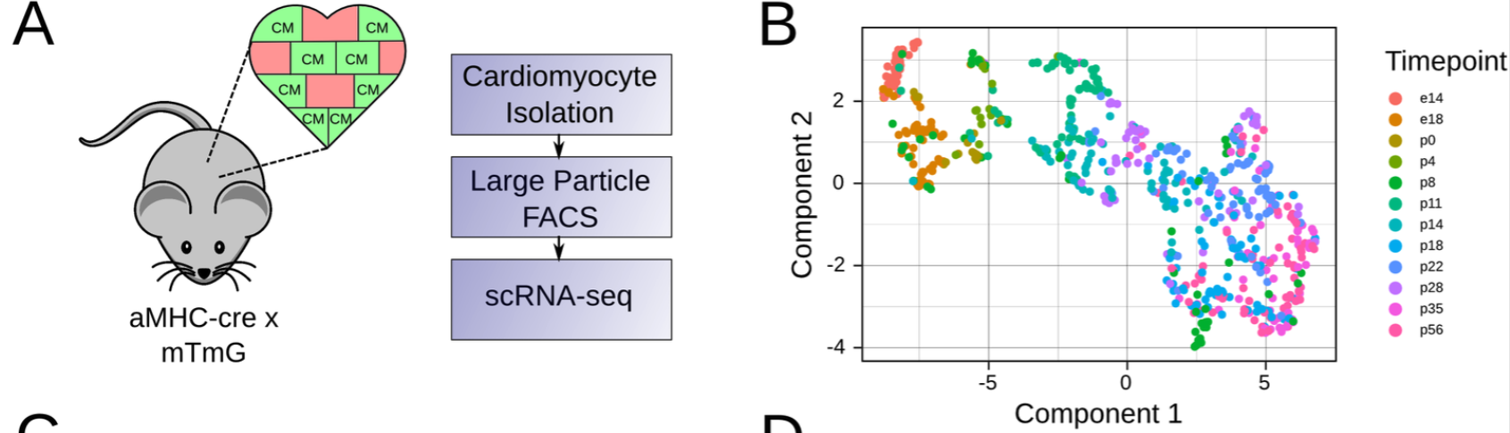
Mouse model used to generate perinatal maturation reference scRNA-seq dataset. B. UMAP of maturation reference dataset. Kannan et al, 2021
Kannan et al have provided a detailed protocol accompanied by software and extensive meta-analysis and validation for their entropy based method. Apply the protocol of Kannan et al. or use some other method to determine the maturation time point that the engineered CMs are most similar to using the perinatal_reference_data. As Kannan et al. do, you should only select the cells that were classified as cardiomyocyte in the previous steps.
What genetic alterations could improve your engineered cardiomyocytes?
Estimating maturity is useful as a metric, but it does not provide an immediate path forward in the form of specific hypotheses for how to improve the engineered CMs. Your collaborators want to identify candidate TFs that they could overexpress or knock out to further enhance differentiation. Set up an in silico screen with CellOracle. The essential components are: 1. A training dataset. To ensure relevant cell types are represented, we recommend the in vivo perinatal CM development timecourse from Kannan et al., available in file folder perinatal_reference_data.
An evaluation dataset. How well do CellOracle’s predicted fold changes correspond to what would actually happen? You can test this using a cardiomyocyte-specific knockout of the transcription factor Prdm16, which was assayed with scRNA-seq at embryonic day 13.5 in figure 6 of Wu et al. 2022.. These data have been downsampled to 5,000 barcodes and are available in the wu_prdm16 folder. There are two biological replicates each of control and knockout samples.
A base GRN. To obtain better performance across a wider range of testable perturbations, compare three options for the base network: completely dense (all possible connections), CellOracle’s default, and a custom base network derived from motif analysis of the ATAC-seq data in Figure 3 of Stone et al, available in direct_conversion_data/GSE130806_Bulk_Atac_Count_Data.bed. As controls, also include randomly generated networks matching the density of the motif-derived networks. There is a tutorial on motif analysis for CellOracle here. If your team has 4 or fewer members, you can skip this item. Just use the default base GRN provided with CellOracle.
A summary metric. Though it’s very useful to have per-gene fold change predictions, they are hard to understand and hard to rank by usefulness. Develop a method to rank the predicted transcriptomes by how useful the perturbations will be for engineering cardiomyocytes.
Look at what TF perturbation would drive the maturation of cardiomyocytes and whether or not it would specifically be beneficial to drive the maturation of cells from directed conversion experiment.
Major Questions (Graduate Section Only):
Option 1:
What non-genetic cues might improve you engineered cardiomyocytes?
Aside from overexpressing transcription factors, another method of optimizing cell fate engineering is to use drug compounds or morphogens that target signaling pathways with important roles in embryonic development. (Box 1 gives some context on morphogens.) Modify or repurpose CellOracle to allow predictions of transcriptomic changes due to activation or repression of signaling pathways. Ideally, any new method of this sort would be tested on a prediction task relevant to in vitro directed differentiation. One potential test dataset is this screen of 320 FDA-approved drugs for maturation of mouse colon organoids, available in the compound_screening folder. In each sample, the full transcriptome is not measured, but roughly 200 genes are measured, which provides enough information to test detailed predictions from an extension of CellOracle. If your solution would benefit from a full-transcriptome intestinal organoid dataset, we suggest the mouse intestinal organoid scRNA-seq dataset from Grün et al. 2015, which uses the same protocol to culture organoids. This dataset is available in the grun_organoids folder.
Box 1: Morphogens. Morphogens are diffusible signals that play a role in patterning of tissues and organs during development. One example of a morphogen is Sonic HedgeHog (SHH). It is a soluble protein. It is important in dorsoventral patterning of vertebrate embryos. During formation of the spinal cord, a flat layer of ectoderm folds inward to form a tube. SHH is secreted from a plate of cells on the innermost side of the tube. It diffuses towards the outermost side. As it reaches the cells that will respond to it, it interacts with membrane-bound protein complexes. Through a moderately complicated series of events, it eventually activates transcription factors from the Gli family. Depending on the dose, different target genes are activated. These target genes repress each others’ expression, giving rise to sharp boundaries between discrete zones with distinct transcriptional profiles. These boundaries remain stable when SHH is removed; the cells are committed to their respective fates. Morphogens provide similar spatial cues throughout development of diverse body structures in diverse organisms.
Morphogen diversity. Morphogens can be soluble proteins like SHH. They can also be small molecules. An example of a natural small-molecule morphogen is retinoic acid, and an example of a natural small-molecule inhibitor of SHH is cyclopamine. (Cyclopamine occurs in plants as a poison, helping defend against herbivorous animals.) Morphogens are often discussed in terms of “pathways”, which describe how the signal reaches the nucleus starting from outside the cell. Other examples of morphogen pathways are Notch, Wnt, bone morphogenic protein (BMP), and transforming growth factor beta (TGF-beta). Each of these has distinct roles, mechanisms, and quirks. There is a surprisingly small collection of pathways that are re-used in many contexts during development of animals with bilateral symmetry. These receive special attention in stem cell biology. But, it is also possible to screen drugs or compounds with no known role in development; they may still prove useful in directed differentiation protocols.
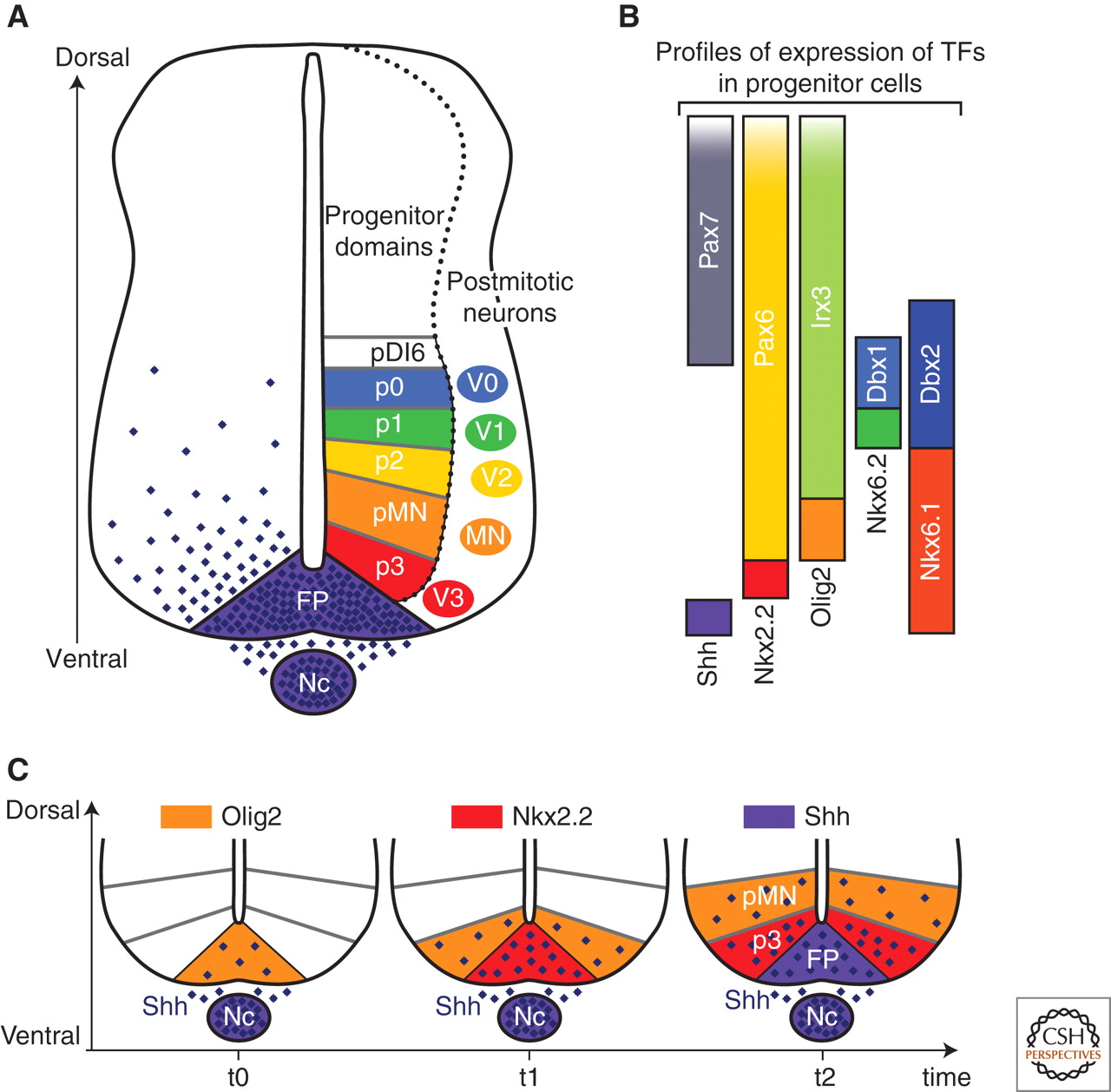
Difference classes of spinal neurons are patterned by different doses of SHH over a fairly short (<24hr) time interval. Image credit: https://cshperspectives.cshlp.org/content/1/2/a002014/F2.expansion.html
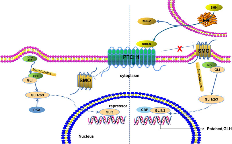
How the SHH signal is transduced into the nucleus to alter transcription.
Image credit: https://www.frontiersin.org/articles/10.3389/fmolb.2021.711710/full
Option 2:
How can your computational methods be translated to help engineer human cardiomyocytes?
Questions 1-3 focus on mouse cells, but your collaborators are ultimately interested in translational and medical research, so they need human CM’s. They have generated preliminary data on directed differentiation of human cells, available in human_engineered_cardiomyocytes (in fact, these data are from Giacomelli et al.). Apply the same assessment and TF screening methods to human iPSCs.